


Board Exam MCQs on Developmental Disturbances of Orofacial Region
Practice. Review mistakes. Master the exam.
How would you like to study?
Start practising
0 correct · 0 incorrect · 0 answered
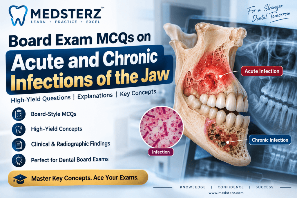
Dental infections involving the jaws are an important topic in oral pathology, oral medicine, oral radiology, and dental board examinations. Questions commonly focus on the differences between acute and chronic infection, osteomyelitis, periosteal reactions, sequestra, radiographic appearances, and characteristic clinical associations.
This post contains board-style MCQs designed to test high-yield concepts related to acute and chronic infections of the jaw. Each question includes five answer choices, the correct answer, and an explanation to reinforce the underlying concept.
Whether you are preparing for NBDE-style examinations, dental postgraduate entrance examinations, or other dental board exams, these questions can help you recognize the clinical and radiographic patterns that are frequently tested.
Topics Covered
In this set of Board Exam MCQs, you will review:
1. Acute Osteomyelitis of the Jaw
- Clinical features of acute jaw infection
- Routes of infection
- Important clinical presentations
- Early radiographic findings
2. Chronic Osteomyelitis
- Chronic infection of the mandibular and maxillary bones
- Sequestrum formation
- Reactive bone formation
- Clinical and radiographic characteristics
3. Garre Osteomyelitis
- Proliferative periostitis
- Occurrence in children and young adults
- Periosteal new bone formation
- Characteristic layered/onion-skin appearance
4. Neonatal Maxillitis
- Acute infection of the neonatal maxilla
- Clinical significance in infants
- Relationship to developing tooth germs
5. Sequestra and Periosteal New Bone Formation
- Identification of sequestra
- Reactive periosteal bone formation
- Imaging evaluation of osteomyelitis
- Role of CT in demonstrating osseous changes
6. Radiographic Findings in Jaw Infections
Important radiographic patterns and terminology, including:
- Inverted J-shaped appearance
- Halo shadow
- Periosteal new bone formation
- Sequestration
- Furcation involvement
7. Furcation Involvement
- Furcation involvement of maxillary molars
- Common routes of periodontal involvement
- Recognition of characteristic radiographic appearances
8. Tuberculosis-Related Findings
- Lupus vulgaris
- Acid-fast bacilli
- Ziehl-Neelsen staining
- Mantoux test interpretation
9. Congenital Syphilis
- Higoumenakis sign
- Skeletal manifestations
- Important board-exam associations
Frequently Asked Questions
What is osteomyelitis of the jaw?
Osteomyelitis is an inflammatory and infectious process involving the bone and bone marrow. In the jaws, it may develop following odontogenic infection, trauma, surgery, or other sources of infection.
What is the difference between acute and chronic osteomyelitis?
Acute osteomyelitis generally represents a recent, active infection and may present with pain, swelling, tenderness, fever, and other signs of acute inflammation. Chronic osteomyelitis persists for a longer period and may be associated with sequestra, sclerosis, draining sinuses, and reactive periosteal bone formation.
What is a sequestrum in osteomyelitis?
A sequestrum is a fragment of devitalized bone that has become separated from the surrounding viable bone. It is an important finding associated with chronic osteomyelitis.
What is Garre osteomyelitis?
Garre osteomyelitis, also called proliferative periostitis, is a chronic inflammatory bone reaction characterized by prominent periosteal new bone formation. It is particularly associated with children and young adults.
Why is Garre osteomyelitis common in young patients?
The periosteum in younger individuals has a strong osteogenic capacity and can respond to chronic irritation or infection by producing substantial amounts of new bone. This contributes to the characteristic periosteal reaction seen in Garre osteomyelitis.
What is neonatal maxillitis?
Neonatal maxillitis is an acute infectious process involving the maxilla in newborns and very young infants. Because of the developing anatomy and tooth germs, infection may have significant consequences.
Which imaging modality is useful for detecting sequestra in jaw osteomyelitis?
CT is particularly useful for evaluating cortical bone destruction, sequestra, and periosteal new bone formation in jaw osteomyelitis.
What is Higoumenakis sign associated with?
Higoumenakis sign refers to unilateral enlargement and thickening of the sternal end of the clavicle. It is classically associated with late congenital syphilis.
What is lupus vulgaris?
Lupus vulgaris is a chronic form of cutaneous tuberculosis.
What does an inverted J-shaped radiographic appearance suggest?
An inverted J-shaped appearance around a maxillary molar can be associated with furcation involvement, particularly when inflammatory bone loss extends toward the furcation region.
What is a halo shadow in relation to jaw infections?
A halo shadow refers to a thin radiographic outline of reactive bone associated with inflammatory pathology adjacent to the maxillary antrum.
What are the important findings to remember for board exams on jaw infections?
High-yield associations include Higoumenakis sign with congenital syphilis, lupus vulgaris with cutaneous tuberculosis, sequestrum with chronic osteomyelitis, proliferative periostitis with Garre osteomyelitis, neonatal maxillitis with acute infection of the neonatal maxilla, and CT for detailed evaluation of sequestra and periosteal new bone.
For example:
- Young patient + periosteal new bone → Garre osteomyelitis
- Sequestrum → chronic osteomyelitis
- Higoumenakis sign → congenital syphilis
- Lupus vulgaris → cutaneous tuberculosis
- Inverted J appearance → consider furcation involvement
- Sequestra + periosteal reaction → CT
Final High-Yield Revision
Before your board examination, make sure you can quickly recognize these associations:
| Finding | Think of |
|---|---|
| Higoumenakis sign | Congenital syphilis |
| Lupus vulgaris | Cutaneous tuberculosis |
| Sequestrum | Chronic osteomyelitis |
| Proliferative periostitis | Garre osteomyelitis |
| Young patient + periosteal new bone | Garre osteomyelitis |
| Neonatal maxillary infection | Neonatal maxillitis |
| Sequestra + periosteal new bone | CT |
| Inverted J-shaped appearance | Furcation involvement |
| Halo shadow near maxillary antrum | Reactive bone formation |
Practice these MCQs carefully and focus on understanding why each answer is correct—not simply memorizing the answer. Board examinations often change the wording or clinical presentation while testing the same underlying concept.